Welcome to the Amira-Avizo Software Use Case Gallery
Below you will find a collection of use cases of our 3D data visualization and analysis software. These use cases include scientific publications, articles, papers, posters, presentations or even videos that show how Amira-Avizo Software is used to address various scientific and industrial research topics.
Use the Domain selector to filter by main application area, and use the Search box to enter keywords related to specific topics you are interested in.
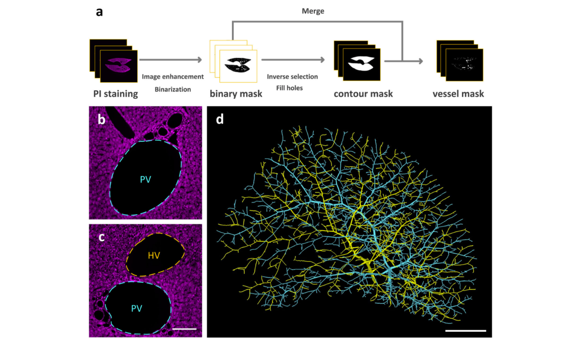
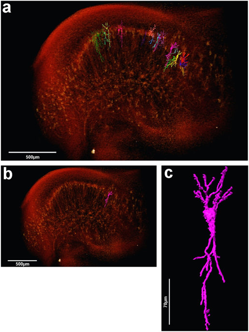
Multiscale reconstruction of various vessels in the intact murine liver lobe
The liver contains a variety of vessels and participates in miscellaneous physiological functions. While past studies generally focused on certain hepatic vessels, we simultaneously obtained all the vessels and cytoarchitectural information of the intact mouse liver lobe at single-cell resolution. […] providing a technology roadmap for studying the fine hepatic vascular structures and their spatial relationship, which will help research into liver diseases and evaluation of medical effi... Read more
Qi Zhang, Anan Li, Siqi Chen, Jing Yuan, Tao Jiang, Xiangning Li, Qingming Luo,Zhao Feng & Hui Gon
High-Resolution Digital Panorama of Multiple Structures in Whole Brain of Alzheimer's Disease Mice
Our study placed emphasis on solving problems in processing high-throughput bright field images and made attempt in developing a method for the extraction and reconstruction of multiple structures. This will facilitate a better understanding of the cerebral anatomical features under the pathological state of AD and shows extensive application prospect in drug efficacy assessment from brain-wide level.
Simultaneously visualizing Amyloid-β (Aβ) plaque with its surrounding brain structu... Read more
Xianzhen Yin, Xiaochuan Zhang, Jingjing Zhang, Weicheng Yang, Xian Sun, Haiyan Zhang, Zhaobing Gao, Hualiang Jiang
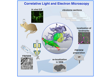
Label-free 3D-CLEM using endogenous tissue landmarks
We demonstrate feasibility of the workflow by combining in vivo 2-photon microscopy and focused ion beam scanning electron microscopy (FIB/SEM) to dissect the role of astrocytic coverage in the persistence of dendritic spines.
Emerging 3D correlative light and electron microscopy (CLEM) approaches enable studying neuronal structure-function relations at unprecedented depth and precision. However, established protocols for the correlation of light and electron micrographs rely ... Read more
Manja Luckner,Steffen Burgold, Severin Filser, Maximilian Scheungrab, Yilmaz Niyaz, Eric Hummel, Gerhard Wanner, Jochen Herms
Insights into data with the KAUST Visualization Core Lab
Through collaboration, the KAUST Visualization Core Lab (KVL) team augments the efforts and domain expertise of KAUST researchers by providing complimentary technical knowledge with exploratory visualization and analytic tools.
KVL’s multi-year collaboration with KAUST Distinguished Professor P. Magistretti and research scientist C. Calì’s KAUST-EPFL Alliance for Integrative Modelling of Brain Energy Metabolism project—itself a collaboration with the Swiss Blue Br... Read more
By the KAUST Visualization Core Lab team and Caitlin Clark
Macroautophagy is morphologically characterized by autophagosome formation. Autophagosomes are double-membraned vesicles that sequester cytoplasmic components for further degradation in the lysosome. Basal autophagy is paramount for intracellular quality control in post-mitotic cells but, surprisingly, the number of autophagosomes in post-mitotic neurons is very low, suggesting that alternative degradative structures could exist in neurons…
Read more
Maria Rosario Fernandez-Fernandez, Desire Ruiz-Garcia, Eva Martin-Solana, Francisco Javier Chichon, Jose L. Carrascosa, Jose-Jesus Fernandez
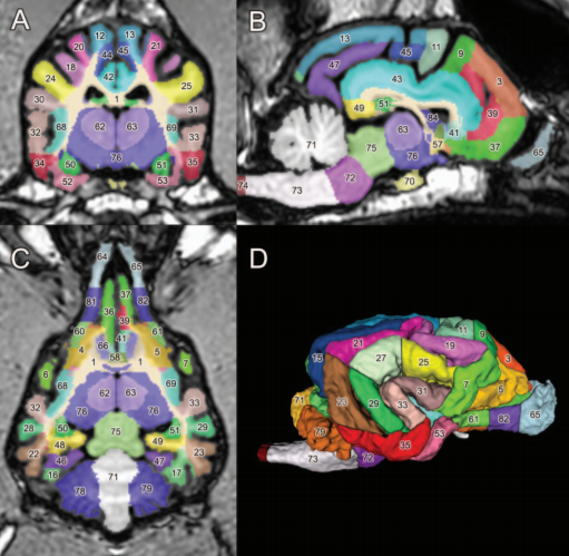
A detailed canine brain label map for neuroimaging analysis
Dogs have recently become an important model species for comparative social and cognitive neuroscience. Brain template-related label maps are essential for functional magnetic resonance imaging (fMRI) data analysis, to localize neural responses. In this study, we present a detailed, individual-based, T1-weighted MRI-based brain label map used in dog neuroimaging analysis. Methods: A typical, medium-headed dog (a 7.5-year-old
male Golden Retriever) was selected from a cohort of ... Read more
Czeibert Kálmán, Andics Attila, Petneházy Örs, Kubinyi Enikő, Kálmán Czeibert
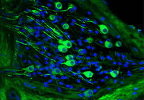
Serotonin (5-hydroxytryptamine, 5-HT) is an important biogenic amine that acts as a neural circuit modulator. It is widespread in the central nervous system of insects. However, little is known about the distribution of serotonin in the nervous system of the cotton bollworm Helicoverpa armigera. In the present study, we performed immunohistochemical experiments with anti-serotonin serum to examine the distribution of serotonin in the central nervous system of H. armigera larv... Read more
Qing-Bo Tang, Wei-Wei Song, Ya-Jun Chang, Gui-Ying Xie, Wen-Bo Chen* and Xin-Cheng Zhao
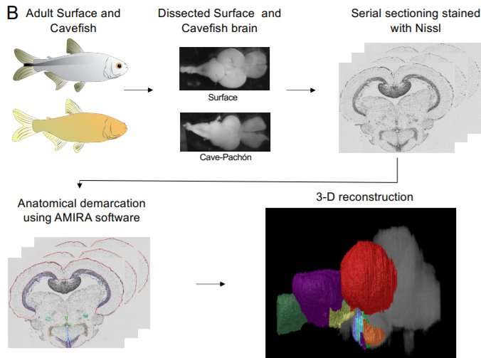
A shift in environmental conditions impacts the evolution of complex developmental and behavioral traits. The Mexican cavefish, Astyanax mexicanus, is a powerful model for examining the evolution of development, physiology, and behavior because multiple cavefish populations can be compared to an extant and ancestral-like surface population of the same species. Many behaviors have diverged in cave populations of A. mexicanus, and previous studies have shown that cavefish ha... Read more
Cody Loomis, View ORCID ProfileRobert Peuß, James Jaggard, Yongfu Wang, Sean McKinney, Stephen Raftopoulos, Austin Raftopoulos, Daniel Whu, Matthew Green, Suzanne E. McGaugh, Nicolas Rohner, Alex C. Keene, Erik R. Duboue
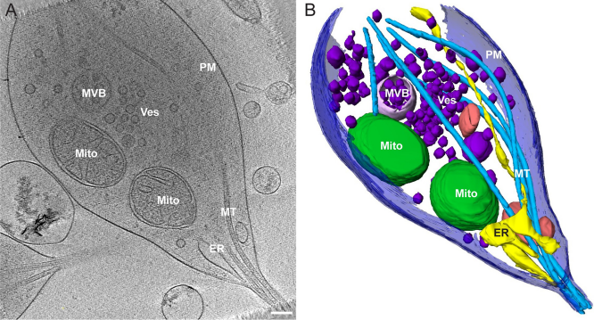
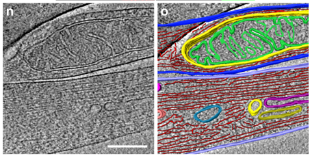
Morphology of mitochondria in spatially restricted axons revealed by cryo-electron tomography
Neurons project axons to local and distal sites and can display heterogeneous morphologies with limited physical dimensions that may influence the structure of large organelles such as mitochondria. Using cryo-electron tomography (cryo-ET), we characterized native environments within axons and presynaptic varicosities to examine whether spatial restrictions within these compartments influence the morphology of mitochondria. Segmented tomographic reconstructions revealed distinctive morphologi... Read more
Tara D. Fischer, Pramod K. Dash, Jun Liu, M. Neal Waxham
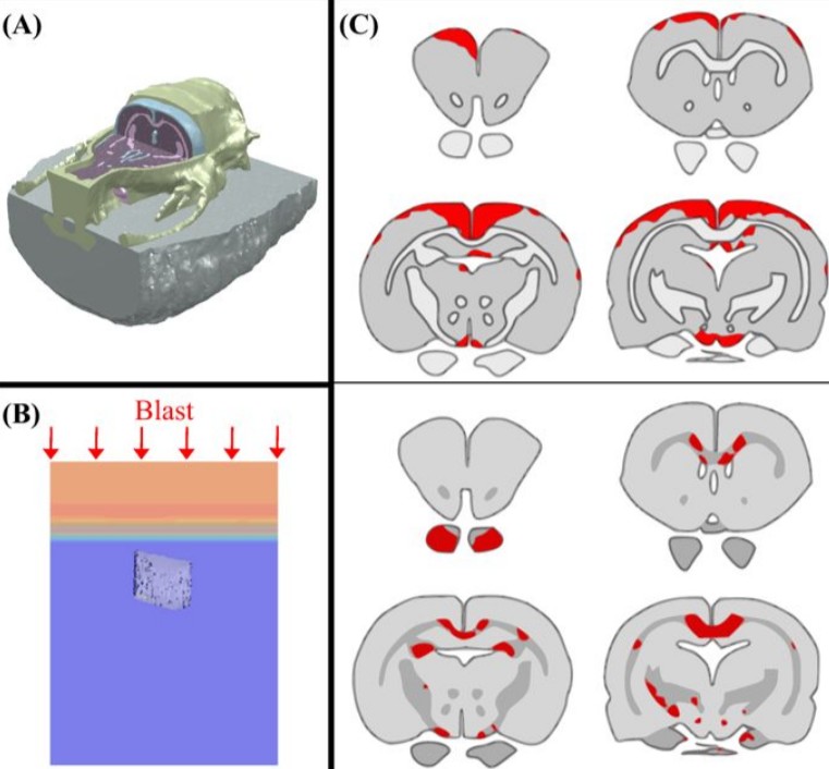
Cognition based bTBI mechanistic criteria; a tool for preventive and therapeutic innovations
Blast-induced traumatic brain injury has been associated with neurodegenerative and neuropsychiatric disorders. To date, although damage due to oxidative stress appears to be important, the specific mechanistic causes of such disorders remain elusive. Here, to determine the mechanical variables governing the tissue damage eventually cascading into cognitive deficits, we performed a study on the mechanics of rat brain under blast conditions. To this end, experiments were carried out to analyse... Read more
Daniel Garcia-Gonzalez, Nicholas S. Race, Natalie L. Voets, Damian R. Jenkins, Stamatios N. Sotiropoulos, Glen Acosta, Marcela Cruz-Haces, Jonathan Tang, Riyi Shi & Antoine Jérusalem
As key functional units in neural circuits, different types of neuronal synapses play distinct roles in brain information processing, learning, and memory. Synaptic abnormalities are believed to underlie various neurological and psychiatric disorders. Here, by combining cryo-electron tomography and cryo-correlative light and electron microscopy, we distinguished intact excitatory and inhibitory synapses of cultured hippocampal neurons, and visualized the in situ 3D organization of ... Read more
Chang-Lu Tao, Yun-Tao Liu, Rong Sun, Bin Zhang, Lei Qi, Sakar Shivakoti, Chong-Li Tian, Peijun Zhang, Pak-Ming Lau, Z. Hong Zhou and Guo-Qiang Bi
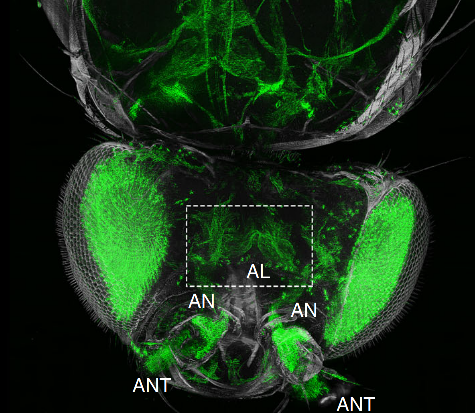
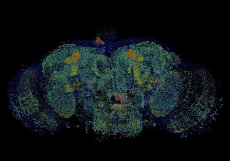
The fruit fly, Drosophila melanogaster, is an important experimental model to address central questions in neuroscience at an organismic level. However, imaging of neural circuits in intact fruit flies is limited due to structural properties of the cuticle. Here we present a novel approach combining tissue clearing, ultramicroscopy, and data analysis that enables the visualisation of neuronal networks with single-cell resolution from the larval stage up to the adult Drosophila. (…) This... Read more
Marko Pende, Klaus Becker, Martina Wanis, Saiedeh Saghafi, Rashmit Kaur, Christian Hahn, Nika Pende, Massih Foroughipour, Thomas Hummel & Hans-Ulrich Dodt
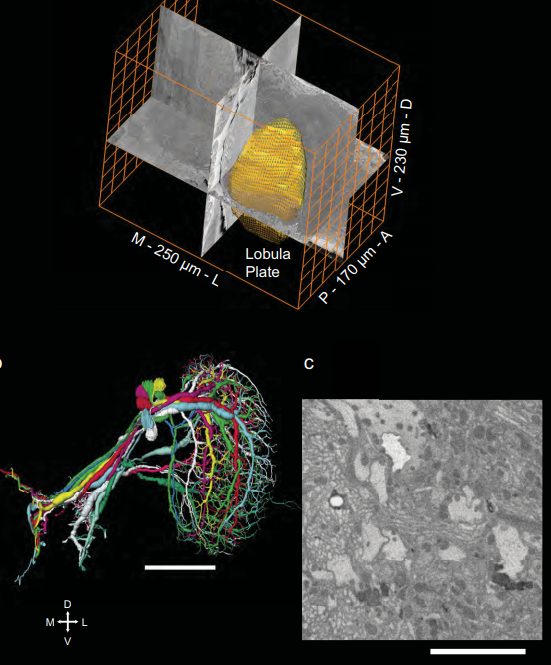
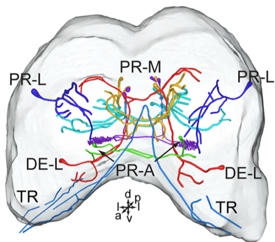
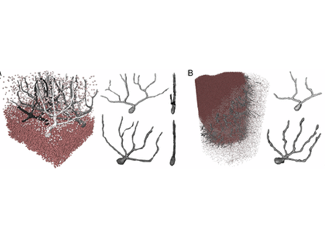
Full reconstruction of large lobula plate tangential cells in Drosophila from a 3D EM dataset
With the advent of neurogenetic methods, the neural basis of behavior is presently being analyzed in more and more detail. This is particularly true for visually driven behavior of Drosophila melanogaster where cell-specific driver lines exist that, depending on the combination with appropriate effector genes, allow for targeted recording, silencing and optogenetic stimulation of individual cell-types. Together with detailed connectomic data of large parts of the fly optic lobe, this has rece... Read more
Kevin M. Boergens , Christoph Kapfer, Moritz Helmstaedter, Winfried Denk, Alexander Borst
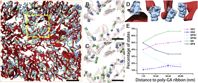
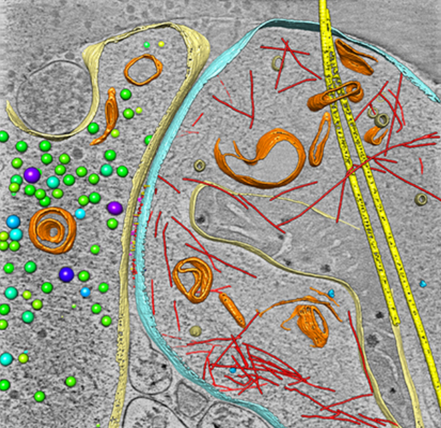
In Situ Structure of Neuronal C9orf72 Poly-GA Aggregates Reveals Proteasome Recruitment
Protein aggregation and dysfunction of the ubiquitin-proteasome system are hallmarks of many neurodegenerative diseases. Here, we address the elusive link between these phenomena by employing cryo-electron tomography to dissect the molecular architecture of protein aggregates within intact neurons at high resolution. We focus on the poly-Gly-Ala (poly-GA) aggregates resulting from aberrant translation of an expanded GGGGCC repeat in C9orf72, the most common genetic cause of amyotrophic latera... Read more
Qiang Guo, Carina Lehmer, Antonio Martinez-Sanchez, Till Rudack, Florian Beck, Hannelore Hartmann, Manuela Perez-Berlanga, Frederic Frottin, Mark S.Hipp, F. Ulrich Hartl, Dieter Edbauer, Wolfgang Baumeister, Ruben Fernandez-Busnadiego
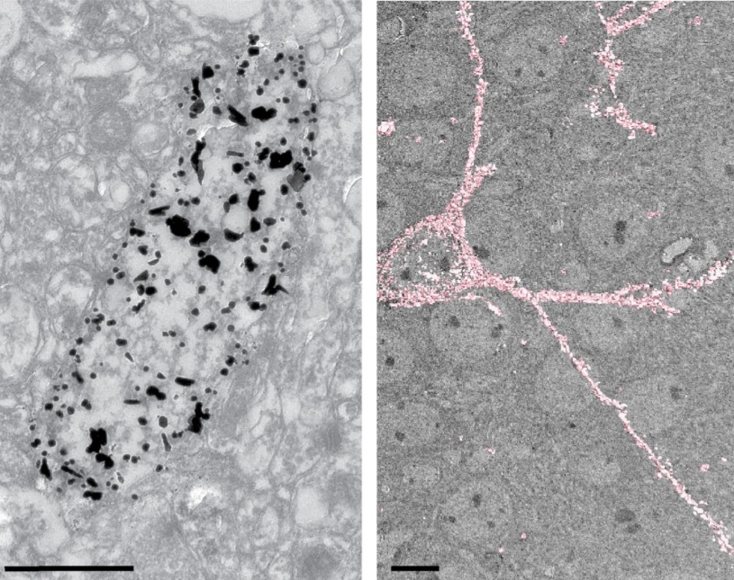
Analysis of neuronal arborization and connections is a powerful tool in fundamental and clinical neuroscience. Changes in neuronal morphology are central to brain development and plasticity and are associated with numerous diseases. Golgi staining is a classical technique based on a deposition of metal precipitate in a random set of neurons. Despite their versatility, Golgi methods have limitations that largely precluded their use in advanced microscopy. We combined Golgi staining with fluore... Read more
Katlijn Vints, Dorien Vandael, Pieter Baatsen, Benjamin Pavie, Frank Vernaillen, Nikky Corthout, Vasily Rybakin, Sebastian Munck & Natalia V. Gounko
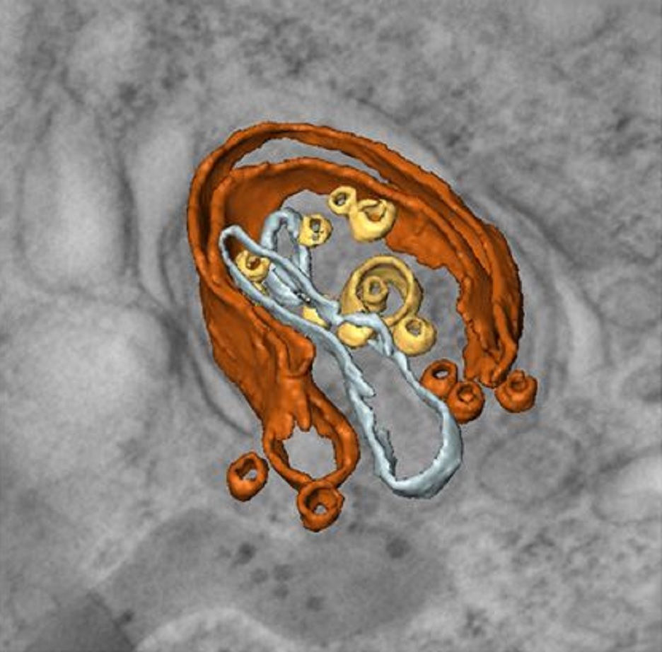
Correlative cryo-electron microscopy reveals the structure of TNTs in neuronal cells
The orchestration of intercellular communication is essential for multicellular organisms. One mechanism by which cells communicate is through long, actin-rich membranous protrusions called tunneling nanotubes (TNTs), which allow the intercellular transport of various cargoes, between the cytoplasm of distant cells in vitro and in vivo. Here, we use correlative FIB-SEM, light- and cryo-electron microscopy approaches to elucidate the structural organization of neuronal TNTs. Our data indicate ... Read more
Anna Sartori-Rupp, Diégo Cordero Cervantes, Anna Pepe, Karine Gousset, Elise Delage, Simon Corroyer-Dulmont, Christine Schmitt, Jacomina Krijnse-Locker & Chiara Zurzolo
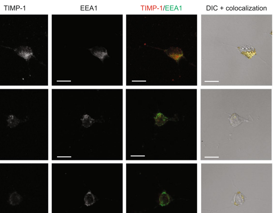
The tissue inhibitor of metalloproteinases-1 (TIMP-1) exerts inhibitory activity against matrix metalloproteinases and cytokine-like effects. We previously showed that TIMP-1 reduces neurite outgrowth in mouse cortical neurons and that this cytokine-like effect depends on TIMP-1 endocytosis mediated by the low-density lipoprotein receptor-related protein-1 (LRP-1). To gain insight into the interaction between TIMP-1 and LRP-1, we considered conformational changes that occur when a ligand bind... Read more
Laurie Verzeaux, Nicolas Belloy, Jessica Thevenard-Devy, Jérôme Devy, Géraldine Ferracci, Laurent Martiny, Stéphane Dedieu, Manuel Dauchez, Hervé Emonard, Nicolas Etique & Emmanuelle Devarenne-Charpentier
The assessment of neuronal number, spatial organization and connectivity is fundamental for a complete understanding of brain function. However, the evaluation of the three-dimensional (3D) brain cytoarchitecture at cellular resolution persists as a great challenge in the field of neuroscience. In this context, X-ray microtomography has shown to be a valuable non-destructive tool for imaging a broad range of samples, from dense materials to soft biological specimens, arisen as a new method fo... Read more
Matheus de Castro Fonseca, Bruno Henrique Silva Araujo, Carlos Sato Baraldi Dias, Nathaly Lopes Archilha, Dionísio Pedro Amorim Neto, Esper Cavalheiro, Harry Westfahl Jr, Antônio José Roque da Silva, Kleber Gomes Franchini
Combined expansion and lattice light sheet microscopy enables high
speed, nanoscale molecular imaging of neural circuits over large volumes.
Optical and electron microscopy have made tremendous inroads in understanding the complexity of the brain, but the former offers insufficient resolution to reveal subcellular details and the latter lacks the throughput and molecular contrast to visualize specific molecular constituents over mm-scale or larger dimensions. We combined expansio... Read more
Ruixuan Gao, Shoh M Asano, Srigokul Upadhyayula, Igor Pisarev, Daniel E Milkie, Tsung-Li Liu, Ved Singh, Austin Graves, Grace H Huynh, Yongxin Zhao, John Bogovic, Jennifer Colonell, Carolyn M Ott, Christopher Zugates, Susan Tappan, Alfredo Rodriguez, Kishore R Mosaliganti, Sean G Megason, Jennifer Lippincott-Schwartz, Adam Hantman, Gerald M Rubin, Tom Kirchhausen, Stephan Saalfeld, Yoshinori Aso, Edward S Boyden, Eric Betzig
Three-dimensional virtual histology of human cerebellum by X-ray phase-contrast tomography
To quantitatively evaluate brain tissue and its corresponding function, knowledge of the 3D cellular distribution is essential. The gold standard to obtain this information is histology, a destructive and labor-intensive technique where the specimen is sliced and examined under a light microscope, providing 3D information at nonisotropic resolution. To overcome the limitations of conventional histology, we use phase-contrast X-ray tomography with optimized optics, reconstruction, and image an... Read more
Mareike Töpperwien, Franziska van der Meer, Christine Stadelmann, and Tim Salditt
Chronic cigarette smoke exposure drives spiral ganglion neuron loss in mice
Tobacco use is associated with an increased risk of hearing loss in older individuals, suggesting cigarette smoke (CS) exposure may target the peripheral auditory organs. However, the effects of CS exposure on general cochlear anatomy have not previously been explored.
Here we compare control and chronic CS exposed cochleae from adult mice to assess changes in structure and cell survival. Two-photon imaging techniques, including the imaging of second harmonic generation (SHG) and two-p... Read more
Stephen T. Paquette, Ryan P. Dawes, Isaac K. Sundar, Irfan Rahman, Edward B. Brown & Patricia M. White